The multi-functional scanner fluorescence imaging system is a high-precision instrument that uses laser precision scanning, time-resolved acquisition and image processing techniques to obtain the photophysical properties of materials in the micro-nano scale space. It is mainly used to study the fluorescence kinetic process and imaging in the micro and nano scale of semiconductor samples, photoelectric conversion materials, photocatalytic materials and biological samples. Its main structure includes laser light source, microscope (ortho or inverted), laser scanning device and detector, etc. It can be fully automated, digital image acquisition and processing through computer control, and can realize various imaging and detection functions such as fluorescence intensity confocal imaging, fluorescence lifetime imaging, carrier migration imaging, micro-nano space fluorescence spectroscopy and Raman spectroscopy acquisition. The system can also be combined with low-temperature device, high-voltage device, (transient) photocurrent/photovoltage detection device and pulse voltage device to achieve a variety of external field conditions of fluorescence kinetics detection, high spatial resolution of photocurrent imaging, electroluminescence kinetics and imaging and other special functions.
01Working schematic

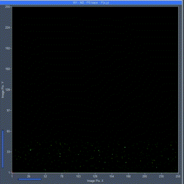
02Software acquisition interface

Achievable functions
- Fluorescence intensity (lifetime) imaging
- Selectable scanning of pixel points
- Settable imaging scan speed
- Spot position programmable
- Adjustable scanning area and magnification
- Fluorescence lifetime auto-fitting
- Fixed-point excitation to observe carrier diffusion
- All data can be exported
03Fluorescence imaging scan video demonstration
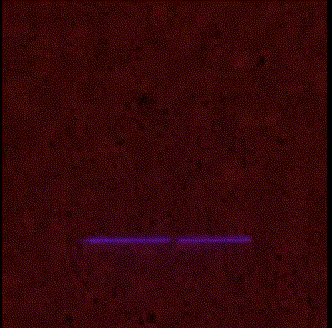
Laser fast scanning video under the microscope Fluorescence imaging scanning video of chalcogenide thin film
(2x accelerated playback) (8x accelerated playback)

Calcium titanite nanowire fluorescence imaging scan video Plant cell fluorescence imaging scan video
(2x accelerated playback) (2x accelerated playback)
04Main technical parameters of the system
I. Laser scanning oscillator module .
1, laser fiber input, distribution control diaphragm system (regulation range: 25μm-2mm)
2, laser scanning imaging range: 256 × 256 pixel dots, up to 4096 × 4096 pixel dots
3、Imaging magnification (zoom): 1-32 times
4、Minimum pixel dwell time: 2.1μs
II. TCSPC Module .
1、Time accuracy: 7 ps
3、Time window: 50 ns - 5 μs
4、Instrument response function (IRF): ≤ 200 ps
5、Spectrum detection range: 400-1000nm
III. Steady-state spectral detection module .
1、Spectrometer (configuration can be adjusted according to customer needs)
2、Focal length: 200mm
3, grating according to the user sample emission wavelength range optional
4、Exit-coupled PMT detector or CCD
2、Spectrum detection mode: wavelength scanning or CCD acquisition
IV. Inverted microscope module .
1, including lighting sources, dichroic, filters and other basic configuration
2, Objective: 100X, 50x, 10x, 5x (according to customer demand optional)
3、Maximum spatial resolution: ~260nm (depending on objective and laser/fluorescence wavelength)
V. Lasers(Optional according to customer needs)
VI. Photocurrent imaging module(Optional according to customer needs)
VII. Electroluminescent module(Optional according to customer needs)
Eight, low temperature / high pressure warehouse module(Optional according to customer needs)
05Application example 1: Fluorescence intensity (lifetime imaging)
Fluorescence intensity and fluorescence lifetime imaging is the basic function of this system, which can perform fast confocal fluorescence imaging of micro-nano-scale samples or sample micro-nano-spatial structures with laser scanning, and can obtain fluorescence intensity and fluorescence lifetime spatial distribution information simultaneously.
Sample: MAPbI3 single crystal nanosheets and MAPbI3 nanowires
Experimental conditions: 100X air microscope, excitation wavelength: 405 nm
Imaging mode: Confocal laser scanning imaging mode

Sample: Calcium titanite polycrystalline film of methylamine lead iodide
Experimental conditions: 100X (oil mirror), excitation wavelength: 405nm
Imaging mode: Confocal laser scanning imaging mode


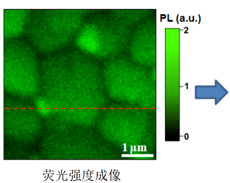

By analyzing the fluorescence intensity change from a to b, the highest spatial resolution up to ~260 nm (the highest spatial resolution reaches the diffraction limit) can be obtained. Effective identification of complex structures such as grain boundaries in chalcogenide polycrystalline films
Samples: Plant cell specimens
Experimental conditions: 50X, excitation wavelength: 405nm
Imaging mode: Confocal laser scanning imaging mode


Sample: 2D SnSe2 (weakly fluorescent material)
Experimental conditions: 100X (oil mirror), excitation wavelength: 405nm
Imaging mode: Confocal laser scanning imaging mode


Enables imaging of fluorescence intensity and lifetime of weakly fluorescent samples
Reference:
Xing Zhou ,et al.,Tianyou Zhai**,Adv. Mater*. 2015, 27, 8035-8041
Source: Tristar Spectrum @ WeChat
Time: 2022/05/10