Expanding Organoid Culture with the MANTIS® Liquid Processor: A Case Study from MIT
Organoids allow researchers to study tissue biology with remarkable fidelity. By using 3D in vitro culture systems to display key features of specific organs, these complex multicellular structures give us physiologically relevant insights that have helped advance many areas of research. A major limitation of traditional organoid culture is that it is often a labor-intensive, high-touch process that requires highly specialized personnel to achieve moderate throughput. Using FORMULATRIX's MANTIS® Liquid Processor, researchers in the Shalek Laboratory at the Massachusetts Institute of Technology (MIT) were able to rapidly screen more than 450 small molecules at varying dosages for their ability to enhance the differentiation of PAN cells within the small intestinal organoids.The MANTIS® provides previously unattainable levels of throughput, and is critical for advancing the understanding of multiple variations in a limited number of samples and materials. materials is critical to advancing the understanding of multiple changes in limited samples.
The intestinal epithelial barrier is an important therapeutic target
Tissue barriers formed by skin, airway and intestinal cells provide interaction with and protection from the environment. They serve to balance body fluids, nutrients, electrolytes, and metabolic waste levels, work closely with the immune system to provide defense against pathogens, and play an important role in tumor surveillance and eradication.
Tissue barrier dysfunction is associated with a wide range of disease states, such as infections, cancers, allergies and various autoimmune diseases. Although environmental exposures can be mitigated with therapies such as antiviral drugs and antibiotics, and immune responses can be modified with vaccines and immunotherapies, in some cases the problem cannot be solved by existing methods.

Despite being a key component of tissue barriers, the intestinal epithelial barrier has been underutilized as a therapeutic target to date. Using organoid systems to model intestinal epithelial barrier tissues from different sources, researchers can better understand these complex systems to develop therapies to treat a wide range of diseases.
Organoid models are driving research progress
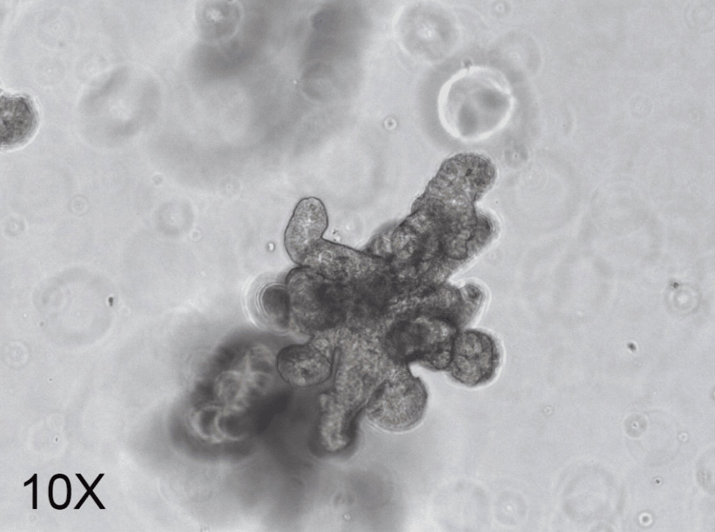
Organoids represent one of the most valuable advances in stem cell research in recent years. Derived from individual adult stem cells, tissue samples containing adult stem cells, or through directed differentiation of pluripotent stem cells, organoids contain populations of stem cells capable of differentiating into organ-specific cell types. These cells exhibit spatial organization and function similar to the organ they represent, giving rise to physiologically relevant systems that mimic in vivo conditions.

Fig. 1. Class organs exhibit spatial organization and function similar to representative organs, such as the crypt/villus morphology of this small intestine class organ.
Organoid studies are limited by flux
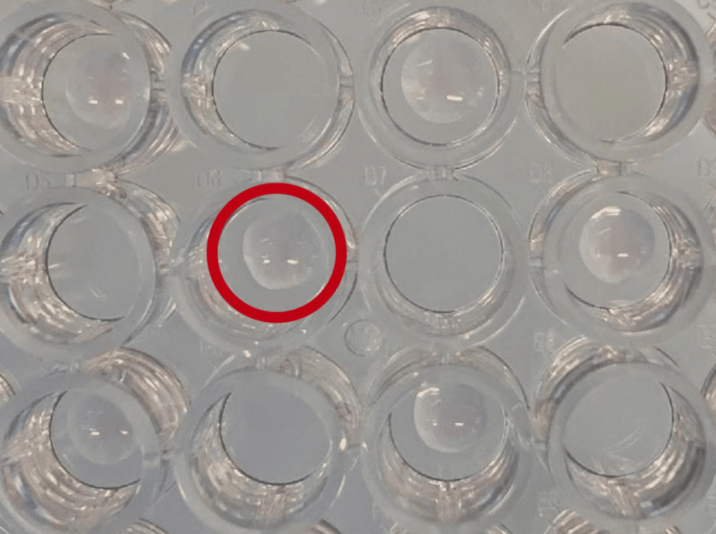
There are a variety of methods for culturing organoids. These include culturing stem cells in the presence of a fibroblast feeder layer or on the surface of a controlled biomaterial matrix, but the most popular method is to encapsulate stem cells in a biologically derived extracellular matrix (ECM) such as Matrigel®. By encapsulating plate-inoculated Matrigel® dome with cell culture medium containing specific growth factors, the cells subsequently proliferate to form three-dimensional structures representative of the investigator's organ of interest.

Figure 2. Matrigel® dome encapsulates stem cells and promotes their proliferation to form physiologically relevant three-dimensional structures.
There are three specific requirements for organoid plate inoculation on Matrigel® dome: 1) precise deposition of cell-loaded ECM onto preheated tissue culture plates, avoiding the edges of the wells to maintain the dome shape necessary for maximum growth; 2) precise manipulation in a very small volume, as sample material is often very limited; and 3) a reasonable degree of temperature control, as the Matrigel® and similar substrates exist as viscous liquids at 4°C and require warm surfaces and environments to form cured hydrogels.
As a result of these requirements, miniaturizing organoid cultures to a scale compatible with conventional screening equipment (96/384/1536 well formats) is extremely challenging. While the rather trivial and time-consuming Matrigel® deposition process can be accomplished manually on 48-well plates, the rate of droplet error formation rises significantly at larger plate volumes and greatly limits the reproducibility of experiments.
MANTIS® is a bench-sized solution for miniaturized organoid research
To overcome the limitations of manual Matrigel® deposition methods, researchers at MIT's Shalek Lab used FORMULATRIX's MANTIS® Liquid Processor to dispense Matrigel® droplets into a variety of plate formats (up to 384 wells). The goal of this study was to accomplish compound screening activities using a small intestinal organoid system at a scale suitable for high-throughput methods.
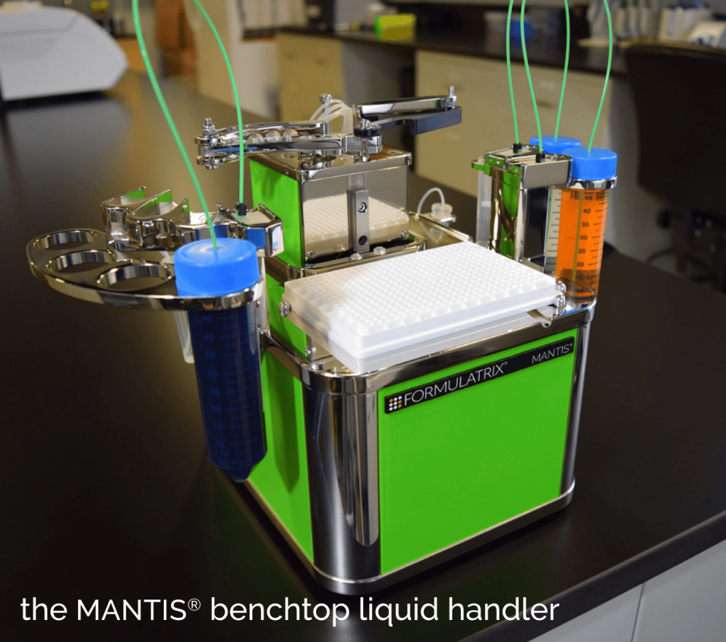
MANTIS® uses a single-channel, non-contact microfluidic dispenser to deliver individual reagents to individual wells one at a time, confining fluids to disposable chips to prevent cross-contamination and eliminating the need for cleaning. An industry-leading CV of less than 31 TP3T supports precise volume delivery down to 0.1 µL while enabling multiple workflow flexibility by accommodating 6-48 chips and handling aqueous solutions up to 25 cP (equivalent to ~601 TP3T of glycerol at room temperature).
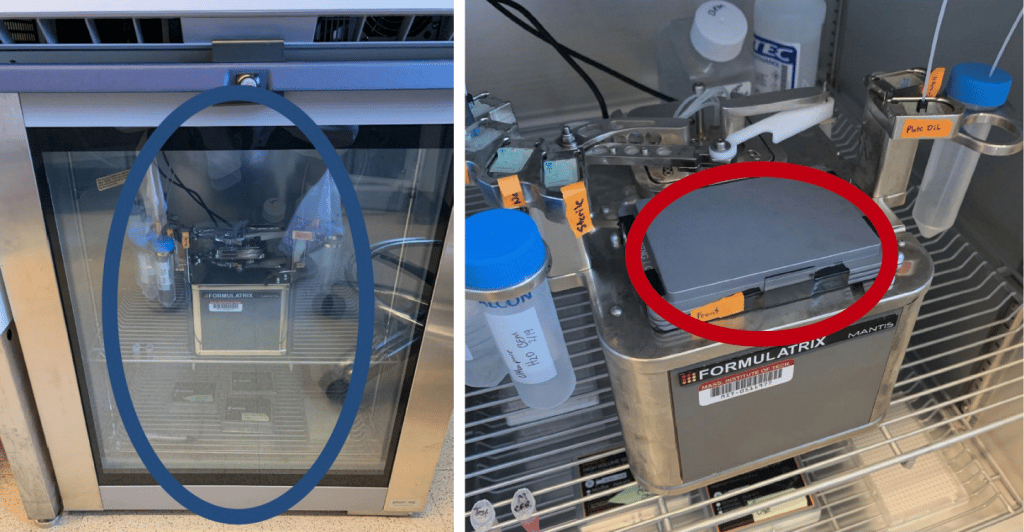
In addition to these features, the MANTIS has a small footprint of only 1ft3, which means it can be easily installed in temperature-controlled environments such as refrigerators or incubators. It is also compatible with a wide range of laboratory software and instruments, enabling seamless integration into existing workflows.

Figure 3. MANTIS® has a very small footprint and can be sealed in a temperature controlled environment. Placing MANTIS® in a refrigerator during this test allowed inoculation of Matrigel® plates onto a heated surface at 4°C (gray = cold, red = warm). These are ideal conditions for the formation of a Matrigel® dome.
Miniaturization of small intestinal organoid cultures gives key insights
Ben Mead, a postdoctoral fellow in MIT's Shalek Laboratory, and colleagues used MANTIS® to miniaturize an existing workflow and screen a library of compounds in a small intestinal organoid system to identify instrumental molecules that may enhance the differentiation of PAN cells. These are the primary anti-microbial producing cells of the human small intestine and are critical for epithelial barrier function.
After applying robust statistical measurements (not yet published), Mead and colleagues identified a number of seedling compounds. Some of these have moved into follow-up studies, providing significant opportunities to depict new biological targets for consideration in the targeted differentiation of PAN cells.
Scaling up increases opportunities for personalized medicine
Several key features of MANTIS® underlie the findings of MIT researchers. By using a single free arm to dispense Matrigel® droplets, MANTIS® provides precise plate inoculation in a variety of plate formats (up to 384 wells) to increase the size of experiments. The instrument can also accommodate 1536-well plates to further expand throughput as needed.
The washable, disposable chip-attached pipette tips allow MIT researchers to handle limited sample volumes while reducing reagent requirements and associated costs. In addition, the small footprint of the MANTIS® means that the entire unit can be easily installed in a standard refrigerator and combined with a pre-warming block to accurately deposit cooled Matrigel® droplets on pre-warmed tissue culture surfaces (see Fig. 4 ); this plate inoculation is a significant improvement over manual techniques.

Figure 4. MANTIS® significantly reduces reagent requirements, including volume of Matrigel® per well, volume of plate inoculation medium, and number of intestinal organoids to be inoculated, as well as reducing plate inoculation time.
Researchers at MIT have demonstrated that the miniaturization approach offered by MANTIS® is effective in studying multiple variations of finite materials. This is relevant for detecting organoids from single patient biopsies and opens up many new opportunities for personalized medicine. As the technology evolves, the inclusion of additional measurement modalities will further improve model fidelity.MANTIS® is the key to realizing the need for larger scale.
Source: @BostonFummerle
Time: 2022.01.11
Related Products
Formulatrix Mantis Microplate Dispenser
Formulatrix USA upgrades micro automated dispensing*
