Cell microarraying (or cell patterning)' is also known as constraining the cells under study to a defined spatial location'It has important potential applications in many areas: The
(1)Drug Development'Using cellular microarrays can significantly reduce the cost of drug development'And more accurate prediction of results;.
(2)Cellular sensors'Microarray technology is an important tool for cell immobilization on cell sensors;
(3)Organizational Engineering'For example, the growth of stem cells into different cell lines depends largely on the shape of the cells'This suggests that surface engineering by controlling the cell growth microenvironment is a means to study functionalized cells;.
(4)Basic Cell Biology'In basic research in molecular biology'Techniques to achieve statute or control of cell adhesion and spreading on the substrate surface'is the key to in-depth studies of cell-substrate and cell-cell interactions.
Cell arrays are generally designed using chemical modifications to form cell-adherent and non-adherent regions on the substrate, and the actual design often requires micromachining of the surface topography or modification of physicochemical properties for different cell types. Commonly used techniques for cell microarraying include:
(1)Photolithography(2)Soft lithography(3)Inkjet technology(4)Stencil-assisted patterning technology(5)Laser Induced Direct Cell Writing Technology(6)Optical fixation and optical construction of microarray technology and(7)Electrochemical patterning technology, etc.
The preparation of cell microarrays by soft lithography will be highlighted here.
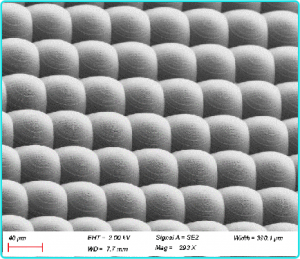
Whitesides et al. prepared cell microarrays by two methods. One was to first dip C18-SH with cellophilic ability using patterned PDMS' and transfer it to the gold film surface 'followed by dipping the gold substrate into a thiol solution containing vinyl glycol terminals (ethylene-glycol-terminated thiols,C11EG3-SH ), which has cell-sparing properties, to form a self-assembled monolayer stopped by EG3 on the remaining gold surface, thus obtaining a surface with selective adhesion to cells. Since the substrate is electrically conductive, after obtaining the cell microarrays, the adsorbed thiols can be desorbed by applying a suitable voltage, so that the cells are no longer immobilized and migrate, thus allowing the study of the patterns of intercellular movement and influence. Another way is to use microfluidic patterning techniques 'using a seal in close contact with the substrate' to introduce a solution containing the molecules to be modified into the trench 'in which these molecules such as the pro-cellular fibrinogen protein are assembled' and after removing the seal Another molecule such as bovine serum albumin (BSA) can be assembled in the region of other unmodified molecules' and a cell-selective substrate can be obtained. The microfluidic chips and cell microarrays obtained using this route are shown in Figure 1.12.
Based on the above work, Grzybowski et al. 'used a chemical wet-stamp technique to etch the Au/glass substrate' to remove part of the Au film and subsequently modified the residual Au island surface with ethylene-glycol- terminated thiols (C11EG3-SH) self-assembled monolayers on the surface of the remaining Au islands' also resulted in a surface with selective adhesion to cells.


This paper is extracted from "Novel electrochemical processing methods for the preparation of three-dimensional micro and nano structures on semiconductor and metal surfaces and their applications", Xiamen University, Tension. For more details, please see the original article

